


by meir vaknin
Copyright © 2023
the body

Copyright © 2023
THE BODY
- Gluteus maximus
- Biceps femoris
- Semitendinosus
- Semimembranosus
- Rectus femoris
- Sartorius
- Tensor fascia latae
- Iliopsoas
- Pectineus
- Gluteus medius and minimus
- Tensor fascia latae
- Piriformis
- Adductor magnus
- Adductor longus
- Adductor gracilis
- Adductor brevis
-
- rectus femoris
- vastus lateralis
- vastus medialis
- vastus intermediusPectineus
- gastrocnemius
- popliteus
- Triceps surae
- gastrocnemius
- soleus
- tibialis posterior
- flexor digitorum longus
- flexor hallucis longus
- peroneus longus and brevis
- tibialis anterior
- extensor hallucis longus
- extensor digitorum longus
- Scalenes (anterior and middle)
- Sternocleidomastoid
- Intercostals (external and internal)
- Diaphragm
- Abdominals:
- Rectus Abdominis
- External Oblique
- Internal Oblique
- Transversus Abdominus
- Erector spinae (sacrospinalis)
- spinalis
- longissimus
- iliocostalis
- Splenius Capitis et cervicis
- Semispinalis
- Multifidus
- Rotatores
- Quadratus Lumborum
- Serratus anterior
- Trapezius
- Rhomboideus major and minor
- Pectoralis minor
- Levator scapulae
- Deltoid
- Rotator cuff
- supraspinatus
- infraspinatus
- teres minor
- subscapularis
- Teres major
- Coracobrachialis
- Latissimus dorsi
- Pectoralis major
- biceps brachii
- brachialis
- brachioradialis
- pronator teres
- triceps brachii
- anconeus
- supinator
- abductor pollicis longus
- extensor pollicis brevis
- extensor indicis proprius
- Radioulnar pronators
- pronator teres
- pronator quadratus
- flexor carpi radialis
- extensor carpi radialis longus
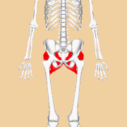
The gluteus maximus
The gluteus maximus is the largest muscle in the human body and plays a significant role in various movements, including hip extension, hip abduction, and hip external rotation. It is located in the buttocks and forms the bulk of the buttock region. Let’s explore the gluteus maximus muscle with the help of some pictures and explanations:
Overview: The gluteus maximus muscle is situated in the posterior (back) aspect of the hip and buttock region. It originates from the posterior part of the iliac crest (the upper curved edge of the hip bone), the sacrum (the triangular bone at the base of the spine), and the coccyx (the small bone at the bottom of the spine). It then inserts into the iliotibial band (a fibrous band of tissue on the outside of the thigh) and the gluteal tuberosity of the femur (the bony prominence on the back of the thigh bone).
Functions: The gluteus maximus muscle has several important functions. It is primarily responsible for extending the hip joint, which means it helps to move the thigh backward. For example, when you push your leg back while walking, running, or climbing stairs, the gluteus maximus contracts to extend the hip. It also assists in hip abduction, which involves moving the thigh away from the midline of the body, and hip external rotation, which involves turning the thigh outward.
Strength and Stability: The gluteus maximus plays a crucial role in providing stability to the hip joint, especially during weight-bearing activities like standing, walking, and running. It helps maintain balance and prevents excessive movement or rotation of the hip joint.
Appearance: The gluteus maximus gives the buttocks their shape and contributes to the overall appearance of the body. Regular exercise and strength training can help tone and strengthen this muscle, leading to a more defined and firm buttock region.
In summary, the gluteus maximus is a powerful muscle located in the buttocks. It is responsible for hip extension, hip abduction, and hip external rotation. This muscle provides strength, stability, and contributes to the appearance of the buttock region. Regular exercise can help keep this muscle strong and well-toned.
Now, let’s take a look at the labeled images:
Image 1: The gluteus maximus as it appears on a skeleton without other muscles
Image 2: The gluteus medius and nearby muscles.
These images should give you a clear visual representation of the Gluteus maximus and its structure.

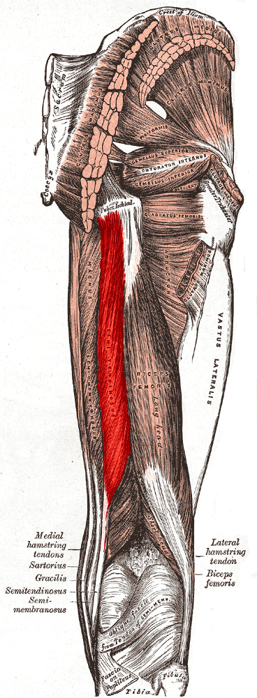
Biceps femoris
The biceps femoris is a muscle located in the back of the thigh. It is one of the muscles that make up the hamstrings muscle group. The biceps femoris plays a significant role in leg extension and hip extension. Let’s explore the biceps femoris muscle in more detail:
Anatomy: The biceps femoris muscle has two heads: the long head and the short head.
- Long Head: The long head of the biceps femoris originates from the ischial tuberosity, a bony prominence in the pelvis. It runs down the back of the thigh, passing behind the knee joint, and inserts onto the head of the fibula, which is the smaller bone of the lower leg.
- Short Head: The short head of the biceps femoris originates from the linea aspera, a ridge on the back of the femur (thigh bone), below the greater trochanter. It also inserts onto the head of the fibula, joining the long head tendon.
Function: The biceps femoris muscle has several functions:
- Leg Flexion: The biceps femoris assists in flexing the knee joint, which involves bending the leg to bring the heel closer to the buttocks.
- Hip Extension: The biceps femoris also helps in extending the hip joint, which means moving the thigh backward.
- External Leg Rotation: The muscle contributes to the external rotation of the lower leg when the knee is flexed.
Strength Training: To strengthen the biceps femoris, exercises that target the hamstrings can be performed. Some common exercises include hamstring curls, Romanian deadlifts, glute bridges, and hip thrusts. These exercises help to develop strength, stability, and overall muscle balance in the hamstring region.
Injuries: The biceps femoris, like other hamstring muscles, can be prone to strains or tears. These injuries often occur during activities that involve sudden acceleration or deceleration, such as sprinting or jumping. Hamstring strains can range from mild to severe and may require rest, rehabilitation, and medical attention depending on the severity of the injury.
Now, let’s take a look at the labeled image:
Image 1: Posterior view of right leg. Long head of muscle highlighted (red), short head (yellow) labeled in the lower part of the image.
These images should give you a clear visual representation of the biceps muscle and its structure.

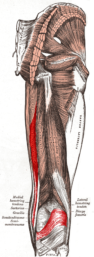
Semitendinosus
The semitendinosus is a muscle located in the back of the thigh, specifically in the hamstrings muscle group. It plays an important role in various leg movements, including knee flexion and hip extension. Let’s explore the semitendinosus muscle in more detail:
Anatomy: The semitendinosus muscle is one of the three muscles that make up the hamstrings muscle group, along with the biceps femoris and semimembranosus muscles. It is a long, thin muscle that runs down the back of the thigh.
Origin and Insertion: The semitendinosus muscle originates from the ischial tuberosity, a bony prominence in the pelvis. It shares this origin point with the biceps femoris muscle. The muscle then travels down the back of the thigh and inserts onto the upper part of the tibia, one of the bones of the lower leg, just below the knee joint.
Function: The semitendinosus muscle has several functions:
- Knee Flexion: It plays a key role in flexing the knee joint, which involves bending the leg to bring the heel closer to the buttocks.
- Hip Extension: The semitendinosus also assists in extending the hip joint, which means moving the thigh backward.
- Internal Leg Rotation: The muscle contributes to the internal rotation of the lower leg when the knee is flexed.
Strength Training: Exercises that target the hamstrings can help strengthen the semitendinosus muscle. Some common exercises include hamstring curls (lying, seated, or standing), Romanian deadlifts, glute bridges, and lunges. These exercises help develop strength, stability, and overall muscle balance in the hamstring region.
Injuries: As with other hamstring muscles, the semitendinosus can be prone to strains or tears. These injuries typically occur during activities that involve explosive movements or sudden changes in speed or direction. Hamstring strains can range from mild to severe and may require rest, rehabilitation, and medical attention depending on the severity of the injury.
Unfortunately, as a text-based AI model, I cannot display or show pictures directly. However, you can easily find labeled diagrams or images of the semitendinosus muscle through an internet search
Now, let’s take a look at the labeled image:
Image 1: Muscles of the gluteal and posterior femoral regions. Semitendinosus labeled at bottom left.
These images should give you a clear visual representation of the biceps muscle and its structure.
Semimembranosus
The semimembranosus is a muscle located in the back of the thigh, specifically in the hamstrings muscle group. It is one of the three muscles that make up the hamstrings, along with the semitendinosus and biceps femoris muscles. The semimembranosus muscle plays a crucial role in leg movements, including knee flexion and hip extension. Let’s explore the semimembranosus muscle in more detail:
Anatomy: The semimembranosus muscle is a long, flat muscle located on the posterior (back) aspect of the thigh. It lies deep to the other two hamstring muscles (biceps femoris and semitendinosus) and is located towards the inner side of the leg.
Origin and Insertion: The semimembranosus muscle originates from the ischial tuberosity, a bony prominence in the pelvis, just like the other hamstrings muscles. It extends down the thigh and attaches to the posterior part of the medial condyle of the tibia, one of the bones in the lower leg.
Function: The semimembranosus muscle has several functions:
- Knee Flexion: It plays a significant role in flexing the knee joint, which involves bending the leg to bring the heel closer to the buttocks.
- Hip Extension: The semimembranosus muscle also assists in extending the hip joint, which means moving the thigh backward.
- Internal Leg Rotation: The muscle contributes to the internal rotation of the lower leg when the knee is flexed.
Strength Training: Exercises that target the hamstrings can help strengthen the semimembranosus muscle. Some common exercises include hamstring curls (lying, seated, or standing), Romanian deadlifts, glute bridges, and lunges. These exercises promote muscle growth, strength, and stability in the hamstring region.
Injuries: Like other hamstring muscles, the semimembranosus can be prone to strains or tears. Hamstring strains often occur during activities involving sudden acceleration, deceleration, or overstretching of the muscle. Rehabilitation, rest, and proper training techniques are essential for recovery and preventing further injury.
Now, let’s take a look at the labeled image:
Image 1: Muscles of the gluteal and posterior femoral regions (semimembranosus labeled at bottom)
These images should give you a clear visual representation of the biceps muscle and its structure.
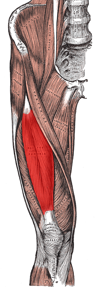
Rectus femoris
The rectus femoris is one of the four muscles that make up the quadriceps muscle group in the front of the thigh. It is the only quadriceps muscle that crosses both the hip and knee joints, making it unique in terms of its function. The rectus femoris plays a vital role in knee extension and hip flexion. Let’s explore the rectus femoris muscle in more detail:
Anatomy: The rectus femoris muscle is a long, strap-like muscle located in the middle of the front thigh. It is the most superficial of the quadriceps muscles and is positioned in the center, with the vastus lateralis on its lateral side and the vastus medialis on its medial side.
Origin and Insertion: The rectus femoris originates from two points:
- Anterior Inferior Iliac Spine (AIIS): It arises from the bony prominence located at the front of the pelvis called the anterior inferior iliac spine.
- Acetabular Labrum: The muscle also has a small attachment to the acetabular labrum, which is a rim of cartilage that surrounds the socket of the hip joint.
From its origin, the rectus femoris extends downward, crossing the front of the hip joint and attaching to the patella (kneecap) through the quadriceps tendon. The quadriceps tendon further continues as the patellar tendon and inserts onto the tibial tuberosity, a bony prominence on the shinbone (tibia).
Function: The rectus femoris muscle has two primary functions:
- Knee Extension: It plays a major role in extending the knee joint, which means straightening the leg from a flexed position.
- Hip Flexion: The rectus femoris is also involved in flexing the hip joint, which means lifting the thigh toward the torso.
Strength Training: To strengthen the rectus femoris, exercises that target the quadriceps can be performed. Some common exercises include squats, leg presses, lunges, leg extensions, and step-ups. These exercises help develop strength, stability, and overall muscle balance in the thigh region.
Injuries: The rectus femoris can be prone to strains or tears, particularly during activities involving explosive movements, sudden changes in speed, or overstretching of the muscle. Rehabilitation, rest, and proper training techniques are crucial for recovery and preventing further injury.
Now, let’s take a look at the labeled image:
Image 1: Muscles of the iliac and anterior femoral regions. (Rectus femoris visible near center.)
These images should give you a clear visual representation of the biceps muscle and its structure.
Sartorius
The Sartorius muscle is the longest muscle in the human body. It is a long, strap-like muscle that runs down the front of the thigh. The Sartorius muscle plays a crucial role in various movements of the hip and knee joints. Let’s explore the Sartorius muscle with the help of pictures and explanations:
Overview: The Sartorius muscle originates from the anterior superior iliac spine (ASIS), which is a bony prominence on the front of the hip bone. It then runs obliquely across the front of the thigh, crossing over the quadriceps muscles. The muscle then inserts into the medial surface of the tibia (shinbone) near the tibial tuberosity.
Functions: The Sartorius muscle has several important functions. It is a multi-joint muscle, meaning it acts on both the hip and knee joints. Its primary function is hip flexion, which involves lifting the thigh toward the abdomen. It also assists in hip abduction, which involves moving the thigh away from the midline of the body. At the knee joint, the Sartorius muscle helps with knee flexion, which is the bending of the knee.
Cross-Legged Sitting: The Sartorius muscle is sometimes referred to as the “tailor’s muscle” because it is actively involved in the cross-legged sitting position. When you sit with one leg crossed over the other, the Sartorius muscle contracts to help flex the hip and externally rotate the thigh.
Injury and Rehabilitation: The Sartorius muscle can be subject to strains or injuries, especially in activities that involve sudden changes in direction or excessive stretching of the muscle. Rehabilitation exercises for Sartorius muscle injuries often focus on strengthening and stretching the muscle to restore its function.
In summary, the Sartorius muscle is a long muscle located in the front of the thigh. It plays a crucial role in hip flexion, hip abduction, and knee flexion. It is involved in movements like sitting cross-legged and is prone to injuries in certain activities. Understanding the function and anatomy of the Sartorius muscle can help in injury prevention and rehabilitation.
Now, let’s take a look at the labeled image:
Image 1: Muscles of the right leg, viewed from the front. (Rectus femoris removed to reveal the vastus intermedius.)
These images should give you a clear visual representation of the biceps muscle and its structure.

Tensor fascia latae
The Tensor fasciae latae (TFL) is a small muscle located on the outer side of the hip. It is part of a group of muscles known as the hip abductors and plays a role in stabilizing the hip joint and controlling the movement of the thigh. Let’s explore the Tensor fasciae latae muscle with the help of pictures and explanations:
Overview: The Tensor fasciae latae muscle is located on the lateral (outer) aspect of the hip. It originates from the anterior iliac crest (the front upper curved edge of the hip bone) and runs down the thigh, attaching to the iliotibial band (a fibrous band of tissue on the outside of the thigh).
Functions: The Tensor fasciae latae muscle has several important functions. Its primary function is hip abduction, which involves moving the thigh away from the midline of the body. It works together with other muscles to stabilize the hip joint during walking, running, and standing on one leg. The TFL also assists in hip flexion and internal rotation.
IT Band Connection: The Tensor fasciae latae muscle connects to the iliotibial band (IT band), a thick band of connective tissue that extends from the hip down to the knee. The IT band plays a role in stabilizing the knee during activities such as walking, running, and jumping.
IT Band Syndrome: The TFL and the IT band can be prone to overuse injuries, leading to a condition called IT band syndrome. This is characterized by pain and inflammation on the outer side of the knee. It is often caused by repetitive activities such as running or cycling, where the IT band rubs against the bony prominence on the outside of the knee joint.
In summary, the Tensor fasciae latae muscle is a small muscle located on the outer side of the hip. It is involved in hip abduction, hip flexion, and hip internal rotation. It works in conjunction with the IT band to stabilize the hip and knee joints. Understanding the function and anatomy of the Tensor fasciae latae muscle can help in addressing issues related to hip and knee stability and injury prevention.
Now, let’s take a look at the labeled image:
Image 1: The tensor fasciae latae and nearby muscles.
These images should give you a clear visual representation of the biceps muscle and its structure.

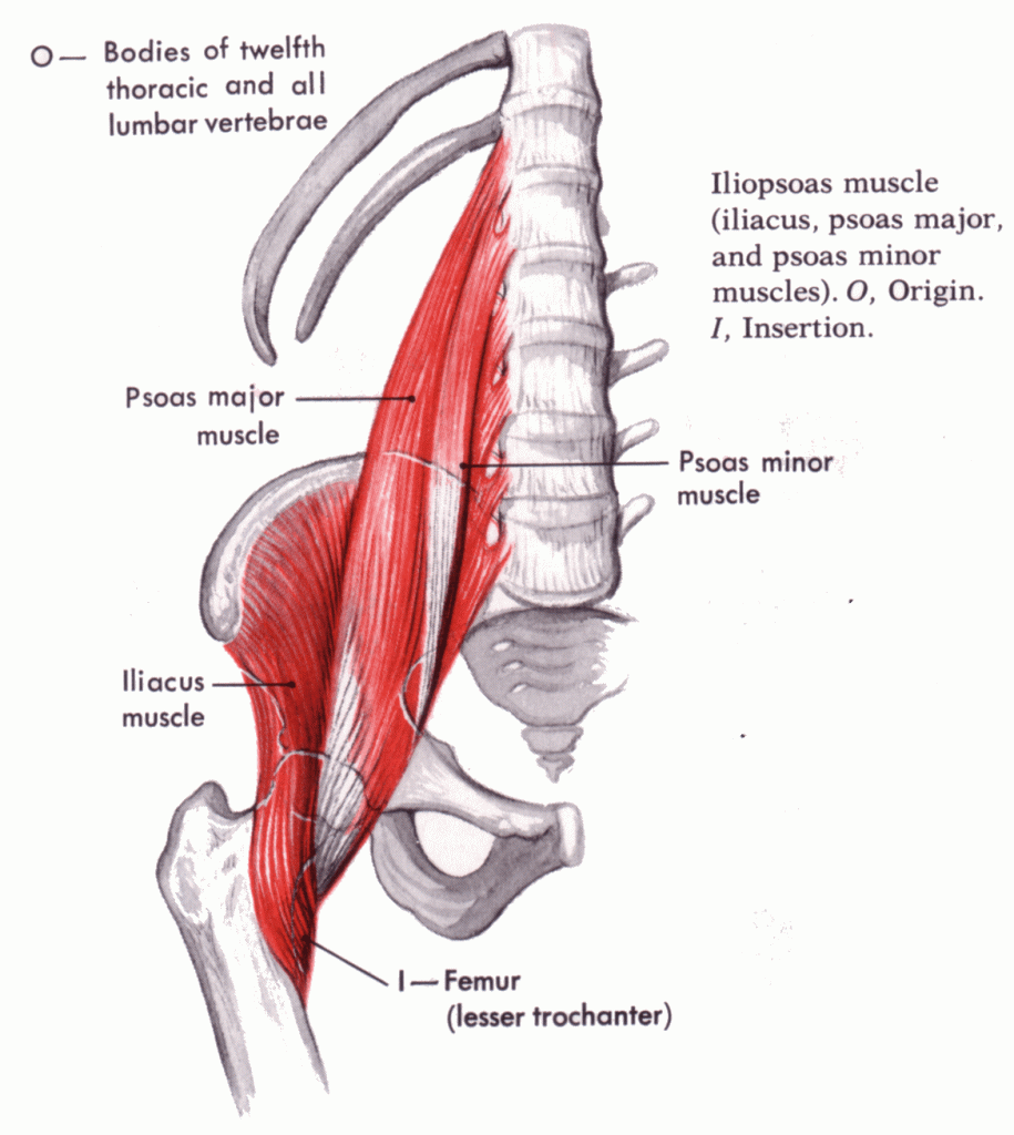
Iliopsoas
The iliopsoas muscle is a group of muscles located in the front of the hip. It consists of two muscles, the psoas major and the iliacus, which work together to flex the hip joint. The iliopsoas muscle is one of the primary muscles involved in activities like walking, running, and climbing stairs. Let’s explore the iliopsoas muscle with the help of pictures and explanations:
Overview: The iliopsoas muscle is a composite muscle formed by the psoas major and iliacus muscles. The psoas major originates from the lumbar vertebrae in the lower back, while the iliacus originates from the iliac fossa, a concave surface on the inside of the hip bone. The two muscles join together and insert into the lesser trochanter, a bony prominence on the femur (thigh bone).
Functions: The iliopsoas muscle has a crucial role in hip flexion, which involves lifting the thigh toward the abdomen. It is the primary muscle responsible for bringing the knee upward when walking or running. Additionally, the iliopsoas muscle assists in stabilizing the lower back and maintaining an upright posture.
Deep Muscle: The iliopsoas muscle lies deep within the abdomen and is not directly visible on the surface. It runs alongside the lower spine, passes through the pelvis, and extends down into the thigh. Due to its deep location, it is not easily accessible for direct palpation or self-massage.
Tightness and Flexibility: The iliopsoas muscle can become tight or shortened due to prolonged sitting, poor posture, or repetitive activities. Tightness in the iliopsoas can lead to discomfort in the lower back, hips, and groin. Regular stretching and flexibility exercises can help maintain the length and flexibility of this muscle.
In summary, the iliopsoas muscle is a composite muscle consisting of the psoas major and iliacus muscles. It plays a vital role in hip flexion and is involved in activities like walking, running, and maintaining posture. Although not directly visible on the surface, the iliopsoas muscle has a deep location within the abdomen. Understanding the function and care of the iliopsoas muscle can help in maintaining hip flexibility and preventing discomfort or injuries in the lower back and hips.
Now, let’s take a look at the labeled image:
Image 1: The Iliopsoas muscle.
These images should give you a clear visual representation of the biceps muscle and its structure.
Pectineus
Gluteus medius and minimus
Tensor fascia latae
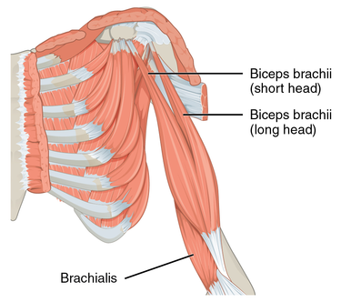
The Biceps Brachii
The biceps brachii is a prominent muscle located in the upper arm. It is a two-headed muscle that plays a significant role in various movements involving the shoulder and elbow joints. Let’s explore the biceps brachii muscle in more detail:
Anatomy: The biceps brachii muscle consists of two heads, the long head and the short head.
- Long Head: The long head of the biceps brachii originates from the supraglenoid tubercle, which is a bony prominence on the scapula (shoulder blade) within the shoulder joint. It runs down the front of the arm and is located more medially (toward the midline of the body).
- Short Head: The short head of the biceps brachii originates from the coracoid process, another bony prominence on the scapula. It is located on the outer side of the arm, more laterally (away from the midline of the body).
Both heads of the biceps brachii merge into a single muscle belly in the upper arm, which then inserts onto the radial tuberosity, a bony prominence on the radius (one of the forearm bones).
Function: The primary function of the biceps brachii is to flex the elbow joint, meaning it brings the forearm closer to the upper arm. This action is commonly known as elbow flexion. Additionally, the biceps brachii also assists in supinating the forearm, which is the motion of rotating the forearm to turn the palm facing upwards. The biceps brachii also has a weak role in flexing the shoulder joint, contributing to arm flexion and raising the arm forward.
Strength Training: The biceps brachii is a popular muscle targeted in strength training exercises due to its visible prominence in the upper arm. Some common exercises that work the biceps brachii include biceps curls, chin-ups, hammer curls, and preacher curls. These exercises involve flexing the elbow against resistance, thereby placing stress on the biceps brachii and promoting muscle growth and strength.
Injuries: The biceps brachii can be subject to various injuries, such as strains, tears, and tendon ruptures. Biceps tendon tears are relatively common and often occur near the attachment points at the shoulder or elbow. These injuries can result from sudden trauma, overuse, or age-related degeneration. Proper warm-up, exercise form, and gradual progression are essential in reducing the risk of injury.
Now, let’s take a look at the labeled images:
Image 1:The biceps is a two-headed muscle and is one of the chief flexors of the forearm. Here is the left side, seen from the front.
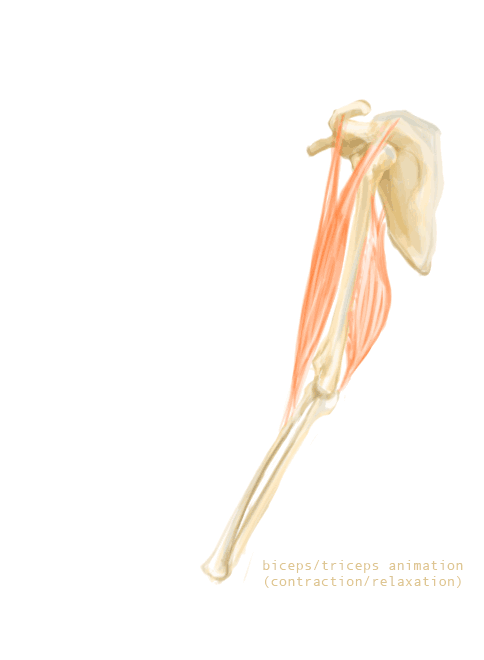
Image 2: Movement of biceps and triceps when arm is flexing.
These images should give you a clear visual representation of the triceps muscle and its structure.
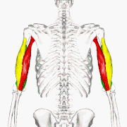
The triceps
The triceps brachii, commonly referred to as the triceps, is a large muscle located on the back of the upper arm. It is the antagonist to the biceps brachii, meaning it performs the opposite action at the elbow joint. The triceps play a crucial role in extending the elbow and are involved in various pushing movements.
Let’s explore the triceps muscle in more detail:
Anatomy: The triceps brachii muscle consists of three heads: the long head, lateral head, and medial head. These heads have different points of origin but converge to form a single tendon that inserts onto the olecranon process of the ulna, the bony prominence at the back of the elbow.
- Long Head: The long head of the triceps originates from the infraglenoid tubercle of the scapula (shoulder blade), below the socket of the shoulder joint. It runs down the back of the arm and is the largest of the three heads.
- Lateral Head: The lateral head arises from the posterior humerus, which is the bone of the upper arm. It is located on the outer side of the arm.
- Medial Head: The medial head originates from the posterior humerus as well, but it lies deeper and closer to the midline of the body.
Function: The primary function of the triceps is to extend the elbow joint, which means straightening the arm from a bent position. This action is commonly known as elbow extension. The triceps are involved in activities such as pushing objects away, performing triceps dips, and stabilizing the arm during certain exercises like bench press or push-ups.
Strength Training: To strengthen the triceps, various exercises can be performed. Some popular exercises include triceps dips, triceps pushdowns, close-grip bench press, overhead triceps extensions, and skull crushers. These exercises involve extending the elbow against resistance, which helps build and tone the triceps muscles.
Injuries: Like any muscle, the triceps can be susceptible to injuries such as strains and tears. However, triceps injuries are relatively less common compared to biceps injuries. Trauma, overuse, or improper lifting techniques can lead to triceps muscle strains or tears. It’s essential to use proper form and gradually increase the intensity of exercises to reduce the risk of injury.
Now, let’s take a look at the labeled images:
Image 1:Long head. Lateral head. Medial head.
Image 2: Movement of biceps and triceps when arm is flexing.
These images should give you a clear visual representation of the triceps muscle and its structure.
Published: Jun 4, 2023
Latest Revision: Jun 4, 2023
Ourboox Unique Identifier: OB-1463356
Copyright © 2023
![]()